Sale Price:
$2,899.99
Original Price:
$2,999.99
The Cellometer Auto 2000 enables users to:
Increase throughput
Increase accuracy
Improve consistency
Count difficult cells (clumpy, irregular-shaped)
Minimize judgment errors, miscounts, interference from red blood cells and user-to-user variability
Additional product information
Primary cell analysis: PBMCs, stem cells, and more
The Cellometer Auto 2000 performs analysis of primary cells from peripheral blood, cord blood, bone marrow, and other complex samples for use in a wide range of research areas, including:
Nucleated cells for transplantation
PBMCs for immunology
Splenocytes for vaccine development
Stem cells for cellular therapy
Tumor cell suspensions for oncology
Dual-color fluorescence allows for staining of live and dead nucleated cells, generating accurate viability results even in the presence of debris, platelets, and red blood cells. Accurate analysis of both ‘messy’ and 'clean' samples enables the Cellometer Auto 2000 to evaluate samples at a variety of points throughout sample processing - from initial collection to separation, to cryopreservation.
Reduced interference from red blood cells, platelets, or debris
The dual-fluorescence AO/PI method utilizes nuclear staining dyes that bind to nucleic acids in the cell nucleus. Because most mature mammalian red blood cells do not contain nuclei, only live and dead mononuclear cells produce a fluorescent signal. There is no need to lyse red blood cells, saving time and removing an extra sample preparation step. Red blood cells, platelets, and debris are not counted in the fluorescent channels.
The advantage of fluorescent counting for primary cells
These images (right) demonstrate the advantage of fluorescent counting for primary cells. The brightfield image shows the combination of nucleated cells, red blood cells, and platelets present in the sample. Only the live and dead nucleated cells are visualized and counted in the green and red fluorescent channels.
 Image 1 of 11
 Image 2 of 11
 Image 3 of 11
 Image 4 of 11
 Image 5 of 11
 Image 6 of 11
 Image 7 of 11
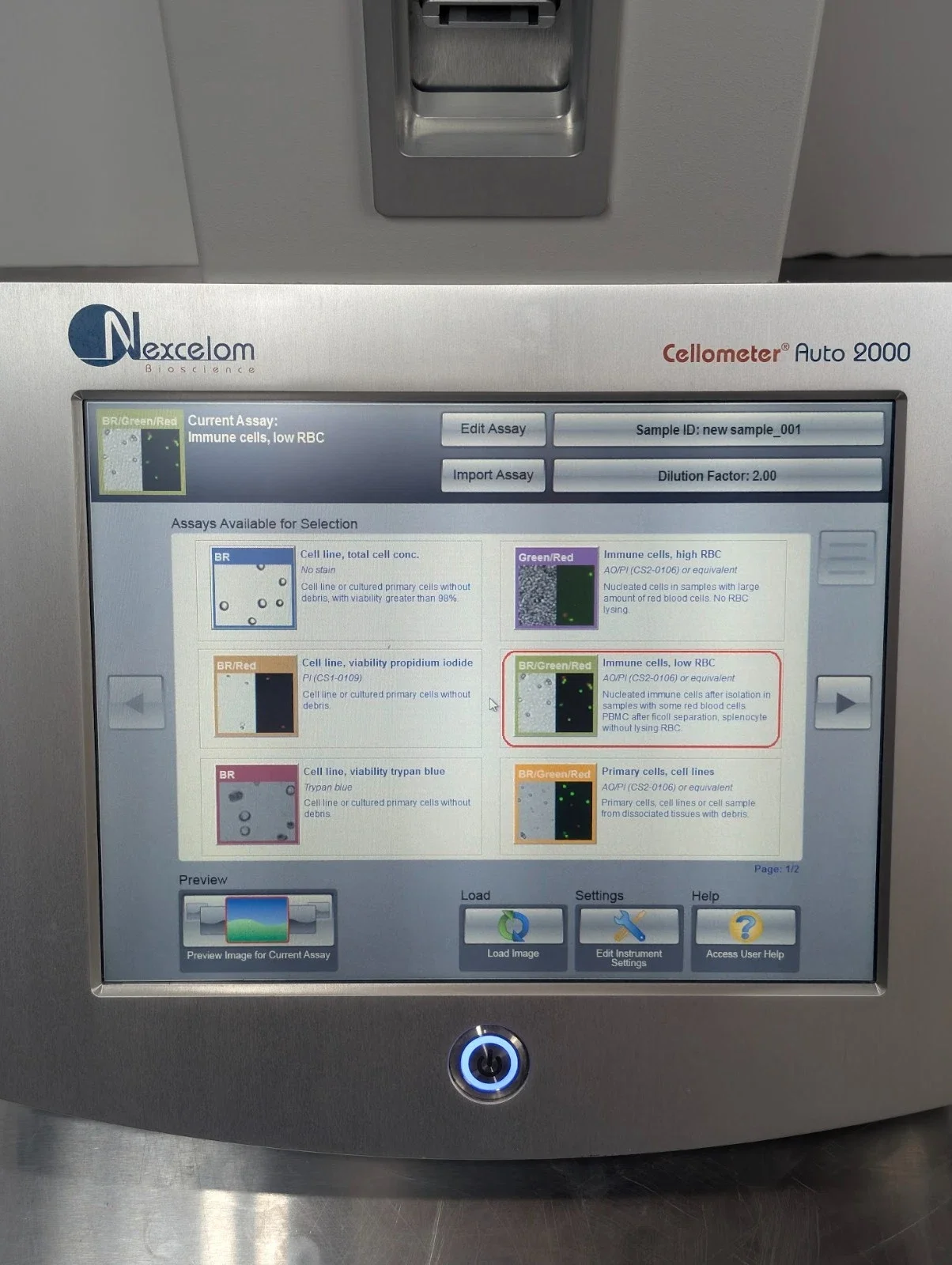 Image 8 of 11
 Image 9 of 11
 Image 10 of 11
 Image 11 of 11